Information magazine of the Department of Industrial Engineering
Università di Trento

Whenever radiation passes through matter, it leaves a kind of “signature” that can be learned to read. Radiation detectors are the instruments that make this possible and are fundamental to a wide range of fields: from high-energy physics experiments, which investigate the fundamental constituents of matter, to medical imaging and clinical dosimetry.
This research activity was carried out as part of a PhD project at the Department of Industrial Engineering (DII), under the supervision of Professors Gian-Franco Dalla Betta and Alberto Quaranta. The work explores two complementary approaches to the development of innovative radiation detectors, both based on the use of new materials and architectures: silicon sensors for neutron detection and scintillators manufactured using 3D printing.
The first research area, conducted within the HYDE2 project, a collaboration between the Italian National Institute for Nuclear Physics (INFN), Fondazione Bruno Kessler (FBK), and the University of Trento (UNITN), focuses on the development of a device for the detection and imaging of thermal neutrons, i.e. low-energy neutrons.
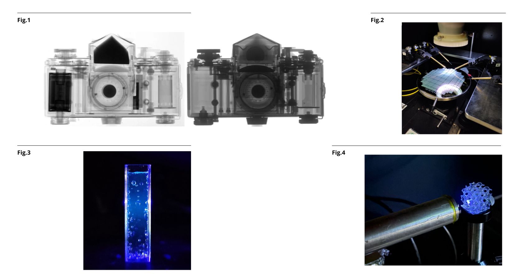
Neutron imaging provides information complementary to that obtained through X-ray imaging, as illustrated in Figure 1. X-rays easily pass through lightweight materials and are more strongly attenuated by dense ones, whereas neutrons interact with matter differently and are particularly sensitive to certain elements, such as hydrogen. This makes neutron imaging a valuable technique for non-destructive testing, with applications ranging from cultural heritage studies and battery inspection to industrial inspections. Since neutrons carry no electric charge, they cannot be detected directly: they must first be converted into charged particles, which in turn generate a measurable signal. The device is based on the use of 3D silicon sensors featuring a microstructured geometry with deep cavities filled with neutron-converter material. This structure increases the probability of neutron capture while maintaining compatibility with pixel-based readout electronics, allowing the acquisition of actual images.
Identifying the optimal cavity geometry represents one of the main design challenges. The device’s operation depends on several interconnected physical processes: from neutron capture to the generation of electrical charges in silicon, followed by their transport and subsequent collection by the electrodes.
To address this challenge, a simulation framework combining three different tools was developed: GEANT4, used to model particle interactions with matter; TCAD Sentaurus, used to simulate semiconductor behaviour; and Allpix², used to reproduce the overall sensor response. This integrated approach makes it possible to efficiently explore a wide range of geometric configurations and accurately estimate detector performance, providing practical guidance for optimizing neutron detection efficiency. The first HYDE devices are already undergoing laboratory testing (Figure 2).
The second research area focuses on scintillators, materials capable of emitting light when exposed to radiation. This phenomenon is related to fluorescence, which can also be observed in everyday life. A familiar example is quinine, found in tonic water, which emits a characteristic blue glow when exposed to ultraviolet (UV) light (Figure 3). The development of these innovative detectors is part of the INFN SHINE project, which aims to develop 3D-printable detectors for applications in hadron therapy, a cancer treatment technique based on proton or ion beams.
Within this project, a new scintillating nanocomposite was characterized. It consists of a polysiloxane-based matrix containing dispersed perovskite nanoparticles, a class of materials that has shown considerable potential as a scintillator in the scientific literature. Its main advantage lies in the possibility of manufacturing the material through stereolithography, a 3D-printing technique that opens the way to detectors with complex geometries, potentially even customized for individual patients. This feature is particularly interesting for clinical dosimetry. Figure 4 shows a 3D-printed SHINE scintillator sample emitting light when exposed to a proton beam. These detectors make it possible to investigate energy deposition in complex geometries, helping to optimize treatments before moving on to characterization directly on patients.
The characterization initially focused on the material’s optical properties, through spectrofluorometric analyses and UV-visible transmittance measurements, to verify that the emitted light is compatible with the readout sensors and that the material provides sufficient optical transparency. Radioluminescence, i.e. light emission induced by radiation, was then investigated using both alpha particles and proton beams at different energies. Some measurements were also performed using therapeutic proton beams in a clinical environment. Comparison with a commercial plastic scintillator showed promising performance.
Although they address different applications, the two research activities share a common goal: developing increasingly efficient, versatile and adaptable detectors to meet the needs of scientific research and clinical applications. On the one hand, the integration of advanced simulations and microstructured silicon technologies makes it possible to design innovative sensors for neutron detection and imaging. On the other, the use of 3D-printable scintillating materials opens up new possibilities for manufacturing devices with customized geometries, particularly promising for dosimetry in hadron therapy.
Two different approaches, both based on the integration of materials engineering, device design and detection technologies, contributing to expanding the possibilities for observing and measuring radiation.
Figure 1: Radiographic images of an analogue camera obtained using neutrons (top) and X-rays (bottom): X-rays highlight the metallic components, while neutrons reveal details of the plastic parts.
Figure 2: HYDE2 devices undergoing testing.
Figure 3: Fluorescence of tonic water under ultraviolet (UV) light.
Figure 4: 3D-printed sample exposed to a proton beam.